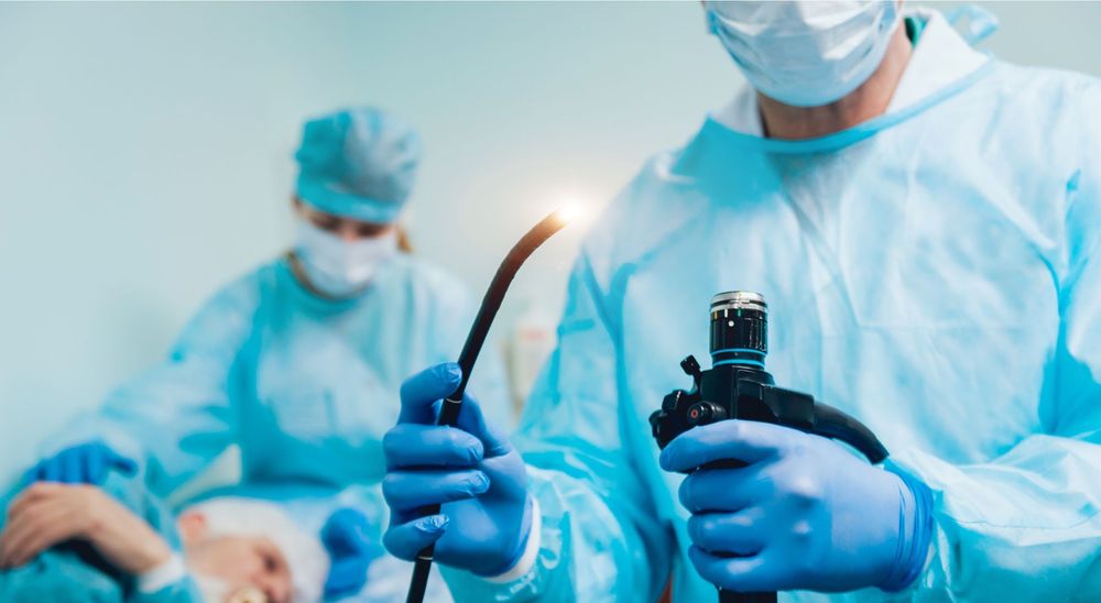
What is EBUS?
EBUS is a minimally invasive procedure that combines bronchoscopy and ultrasound imaging to visualize and access structures deep within the chest, particularly the lungs and mediastinal lymph nodes. Using a special bronchoscope equipped with an ultrasound probe, doctors can accurately locate and biopsy suspicious areas — without the need for surgical cuts.
Why is EBUS Important?
EBUS has revolutionized the diagnosis and staging of lung cancer, infections, and other chest diseases.
Its key benefits include:
✅ High accuracy in detecting cancerous lymph nodes or masses.
✅ No external incision – performed through the airway.
✅ Quicker recovery with minimal discomfort.
✅ Real-time ultrasound guidance ensures precise needle placement.
✅ Outpatient procedure – patients usually go home the same day.
Conditions Diagnosed Using EBUS
EBUS is used to evaluate:
Lung cancer or metastatic disease
Tuberculosis or other chest infections
Sarcoidosis and inflammatory conditions
Enlarged lymph nodes in the chest
How the Procedure is Done
The patient is usually sedated or under light anesthesia. A thin flexible bronchoscope fitted with an ultrasound probe is gently inserted through the mouth into the airways. The ultrasound helps identify the exact location for sample collection. A fine needle is then guided through the airway wall to collect tissue samples for analysis.
EBUS at Arion Radiotherapy & Oncology Centre
At Arion Radiotherapy and Oncology Centre, Bangalore, EBUS procedures are performed by skilled interventional pulmonologists using state-of-the-art ultrasound bronchoscopy systems. Our focus is on accurate diagnosis, patient comfort, and early detection — helping clinicians design personalized treatment plans for every patient.
Conclusion
EBUS has transformed the way lung and chest conditions are diagnosed — making it safer, quicker, and more reliable. If you or a loved one are advised a chest biopsy or evaluation for lung cancer, EBUS is the most advanced and minimally invasive option available.